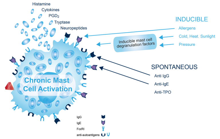
The central role of the mast cell as primary effector cell in chronic urticaria is well established. Chronic spontaneous urcitaria or inducible urticaria are caused by the inappropriate activation and degranulation of dermal mast cells. Mast cells are located in the upper dermis, the ideal situation for wheal formation and sensory nerve stimulation. Increased numbers of mast cells are found in both lesional and non‐lesional skin in CSU and inducible urticaria.
Various activating factors stimulate mast cells to secrete vasoactive molecules that activate endothelial cells leading to increased vasopermeability and extravascular leakage of fluids and proteins (urticaria wheals). Mast cell degranulation in the area of wheals has been demonstrated repeatedly by light and electron microscopy. Histamine, PGD2, tryptase and further mast cell cytokines and neuropeptides, such as nerve growth factor, directly and indirectly (e.g. Th2 type inflammation) drive the inflammation leading to chronic urticaria and the related symptoms such as dermal whealing – both PGD2 and tryptase being quite specific for mast cells.
The global prevalence for chronic urticaria is estimated at about 1% to 1,2% with an estimated 70 million patients globally.
SCIENTIFIC SOURCES
- Church M. K., Kolkhir P., Metz M., Maurer M.: The role and relevance of mast cells in urticarial, Immunol Rev. 2018 Mar;282(1):232-247.
- Jain S.: Pathogenesis of chronic urticaria: An overview, dermatology research and practice 2014;2014:674709
- Clive E.H. Grattan and Elena Borzova: 42 – urticaria, angioedema, and anaphylaxis, editor(s): Robert R. Rich, Thomas A. Fleisher, William T. Shearer, Harry W. Schroeder, Anthony J. Frew, Cornelia M. Weyand, clinical immunology (Fifth Edition), 2019, Pages 585-600